People
Table view of Current People
| Name | Position | Links | |
|---|---|---|---|
| Akshaye Pal | PhD Student | apal@mpi-cbg.de | |
| Alberto Hernandez-Armendariz | Postdoc | ahernand@mpi-cbg.de | |
| Andrea Zinke | Technician | zinke@mpi-cbg.de | |
| Andrey Pozniakovsky | Staff Scientist | andrei.pozniakovsky@mpi-cbg.de | |
| Anne Schwager | Technician | schwager@mpi-cbg.de | |
| Anupa Majumdar | Postdoc | majumdar@mpi-cbg.de | |
| Carsten Hoege | Research Scientist | hoege@mpi-cbg.de | |
| David Kuster | PhD Student | kuster@mpi-cbg.de | |
| Enikö Kovács | Assistant to Tony | kovacs@mpi-cbg.de | |
| Iris Smokers | Postdoc | smokers@mpi-cbg.de | |
| Jon Savage | Postdoc | savage@mpi-cbg.de | |
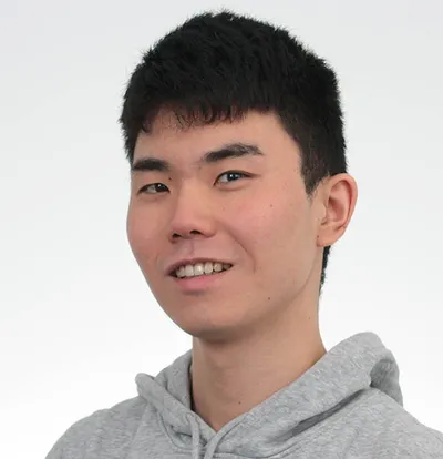
| Koichiro Takenaka | PhD Student | takenaka@mpi-cbg.de | |
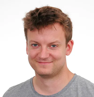
| Lars Hubatsch | Postdoc | hubatsch@mpi-cbg.de | |
| Meline Macher | Postdoc | macher@mpi-cbg.de | |
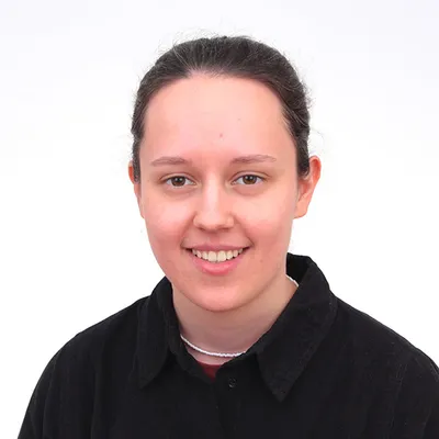
| Silja Zedlitz | PhD Student | zedlitz@mpi-cbg.de | |
| Smitaroopa Kahali | Postdoc | ||
| Susanne Ernst | Technician | ernst@mpi-cbg.de | |
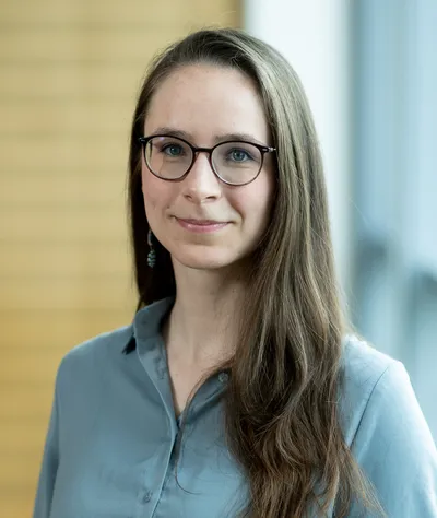
| Theresia Gutmann | Scientist | theresia.gutmann@embl.de | |
| Xiao Yan | Postdoc | xyan@mpi-cbg.de | |
| Yu (Elsa) Wei | PhD Student | wei@mpi-cbg.de | |
| Yuri Hong | Postdoc | hong@mpi-cbg.de |
Current People
-
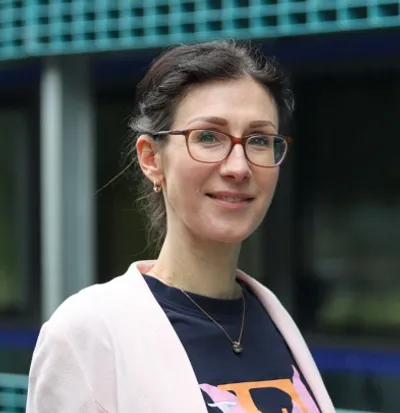
Enikö Kovács
Assistant to Tony -
 © MPI-CBG
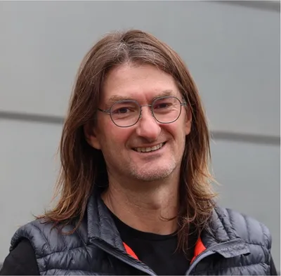
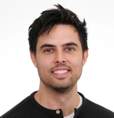
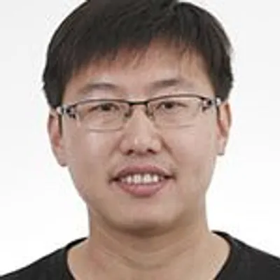
Carsten Hoege
Research Scientist -
 © MPI-CBG
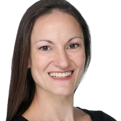
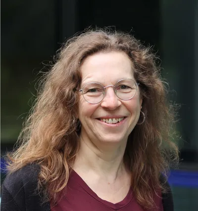
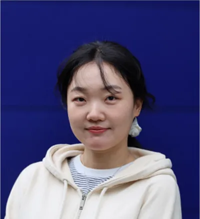
Theresia Gutmann
Scientist -
 © MPI-CBG
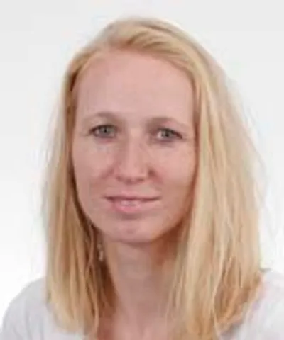
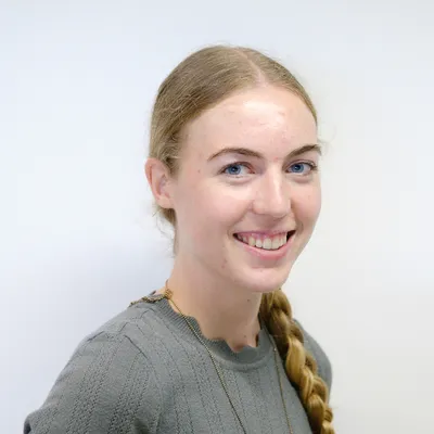
Andrea Zinke
Technician -
 © MPI-CBG
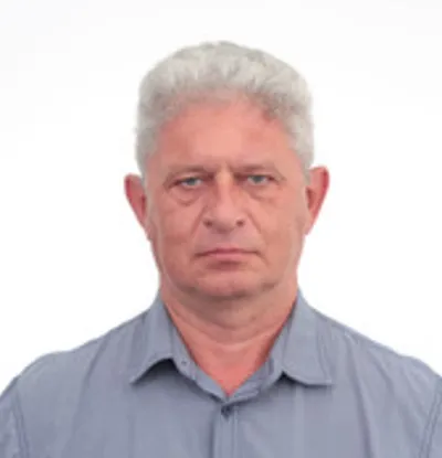
Andrey Pozniakovsky
Staff Scientist -
 © MPI-CBG
Anne Schwager
Technician -
 © MPI-CBG
Susanne Ernst
Technician -
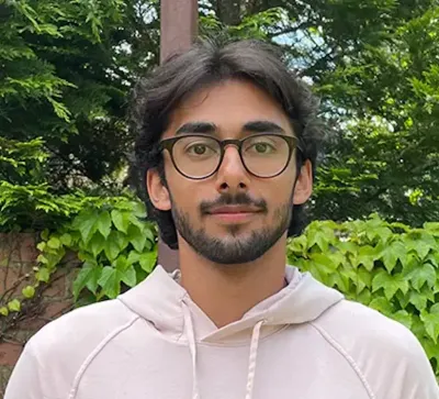
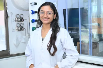
Akshaye Pal
PhD StudentJoint student with the Zechner lab -
 © MPI-CBG
Alberto Hernandez-Armendariz
PostdocGuest scientist from Ebisuya-Lab -
 © MPI-CBG
Anupa Majumdar
Postdoc -
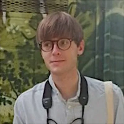
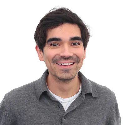
David Kuster
PhD Student -
Iris Smokers
Postdoc -
 © MPI-CBG
Jon Savage
Postdoc -
 © MPI-CBG
Koichiro Takenaka
PhD Student -
 © MPI-CBG
Lars Hubatsch
Postdoc -
Meline Macher
Postdoc -
 © MPI-CBG
Silja Zedlitz
PhD Student -
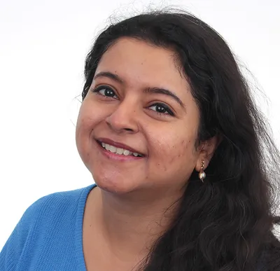
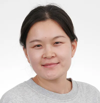
Smitaroopa Kahali
Postdoc -
 © MPI-CBG
Xiao Yan
Postdoc -
 © MPI-CBG
Yu (Elsa) Wei
PhD Student -
Yuri Hong
Postdoc